
Cells Haleo
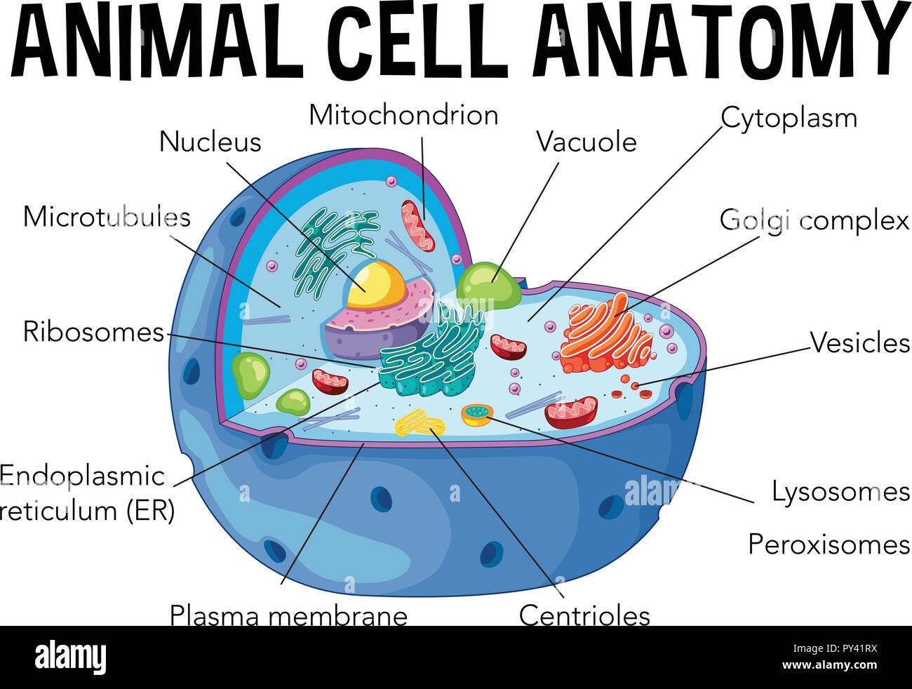
A Labeled Diagram of the Animal Cell and its Organelles There are two types of cells - Prokaryotic and Eucaryotic. Eukaryotic cells are larger, more complex, and have evolved more recently than prokaryotes. Where, prokaryotes are just bacteria and archaea, eukaryotes are literally everything else.
Questions And Answers On Labeled/Unlebled Diagrams Of A Human Cell Human Cell Diagrams Labeled
Structure and Components of a Human Cell. Cell is a compartment where all the activities of life takes place. There are two basic types of cells in nature, viz., prokaryotic cells and eukaryotic cells. Comparison of Prokaryotic Cells and Eukaryotic Cells: Prokaryotic Cells: ADVERTISEMENTS: 1.

Parts of a Cell
Cells are the fundamental unit of life. All living things are composed of cells. While there are several characteristics that are common to all cells, such as the presence of a cell membrane, cytoplasm, DNA and ribosomes, not all cells are the same. Prokaryotic cells lack a nucleus and membrane-bound organelles.
Cell Biology, Cell Structure
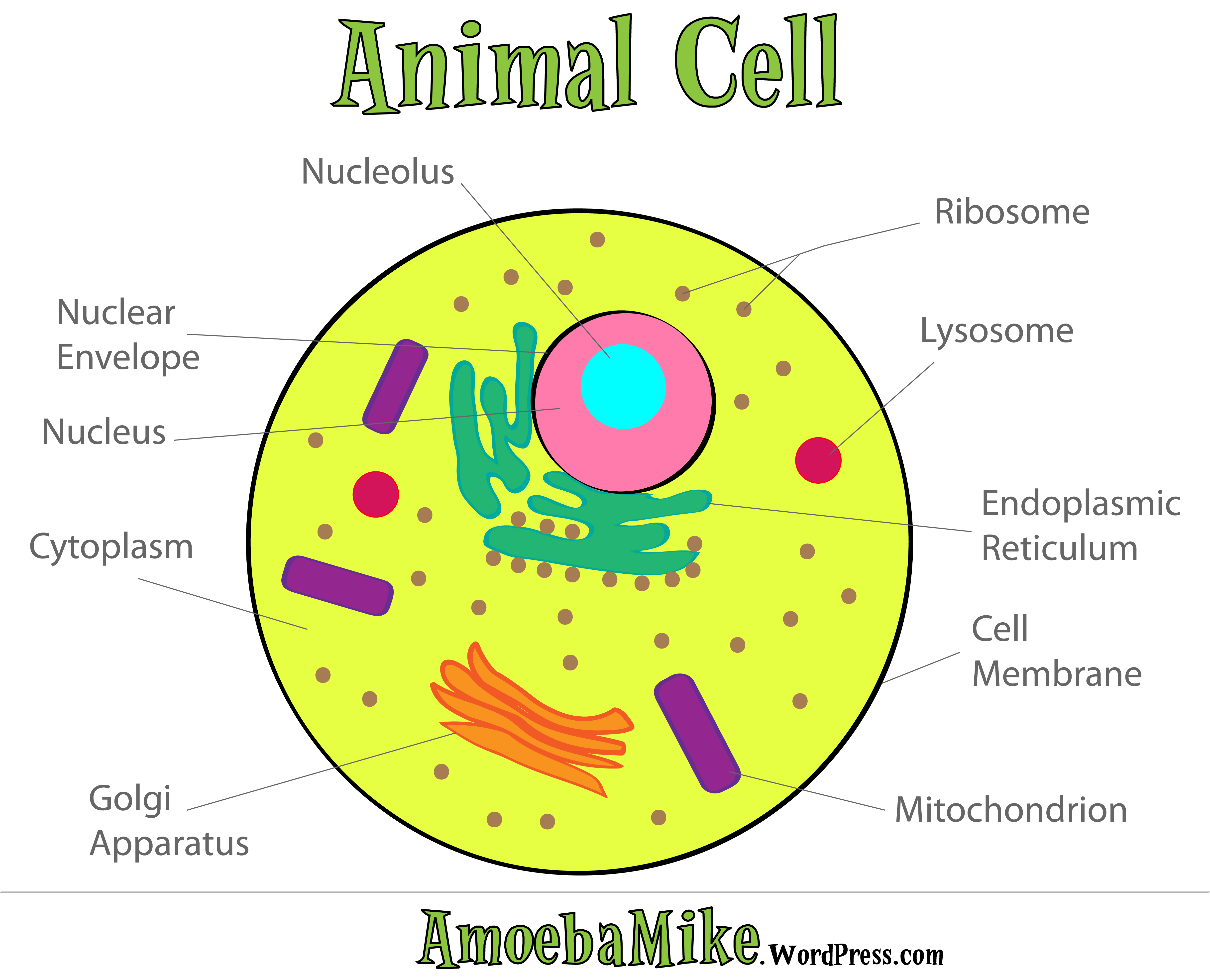
Animal Cell Diagram, Structure, Types, Functions. Definition of animal cell, Animal cell size and shape. Animal cell are considered to be the fundamental living species belonging to the kingdom Animalia. They are eukaryotic cells which means they possess an actual nucleus as well as organelles, which are special structures which perform various functions.

Cell Anatomy Vector Illustration Labeled Educational Structure Diagram Stock Illustration
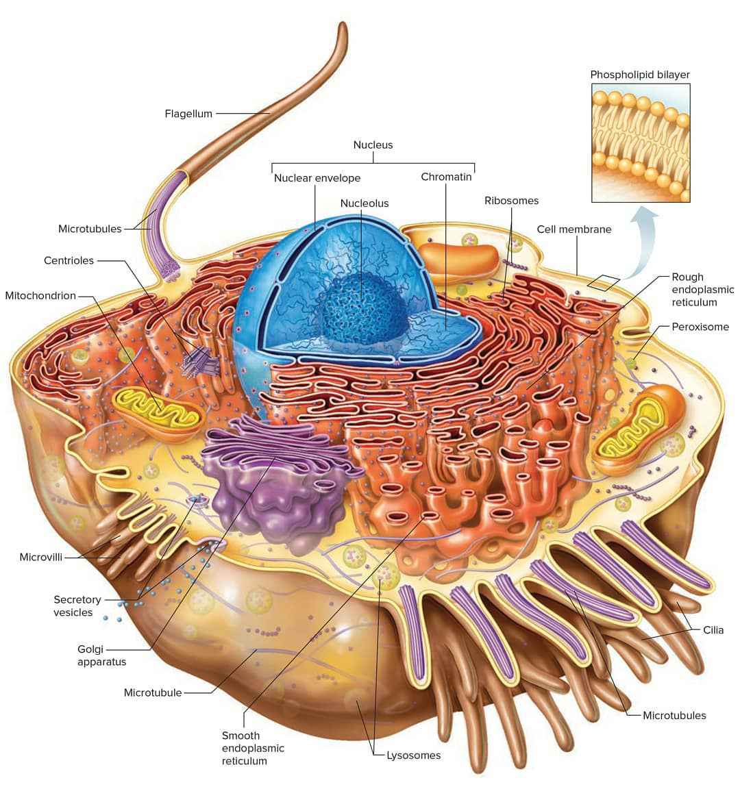
What are the key features of eukaryotic cells? Unlike prokaryotic cells, eukaryotic cells have: A membrane-bound nucleus, a central cavity surrounded by membrane that houses the cell's genetic material. A number of membrane-bound organelles, compartments with specialized functions that float in the cytosol.

Cell Structure
cell, in biology, the basic membrane-bound unit that contains the fundamental molecules of life and of which all living things are composed. A single cell is often a complete organism in itself, such as a bacterium or yeast. Other cells acquire specialized functions as they mature. These cells cooperate with other specialized cells and become.

Explain the nucleus of a cell with a neat labeled diagram Science Cell Structure and
What exactly is its job? The plasma membrane not only defines the borders of the cell, but also allows the cell to interact with its environment in a controlled way. Cells must be able to exclude, take in, and excrete various substances, all in specific amounts.
The Cell AmoebaMike
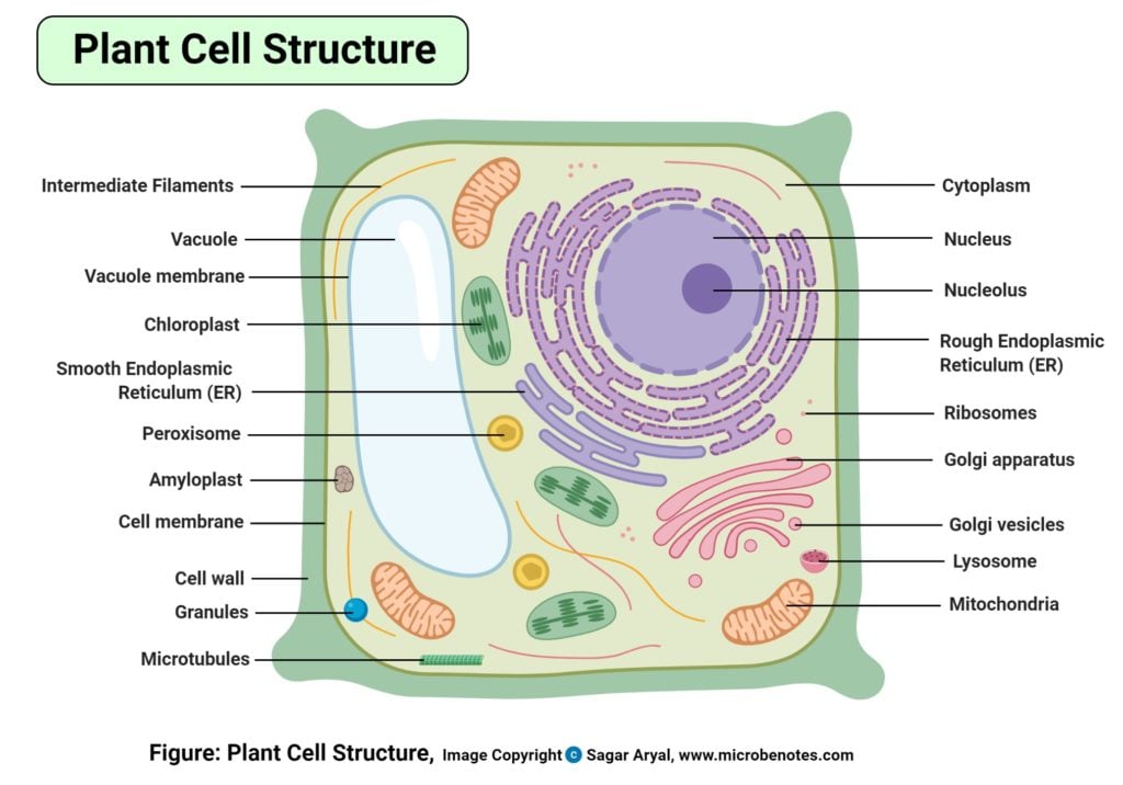
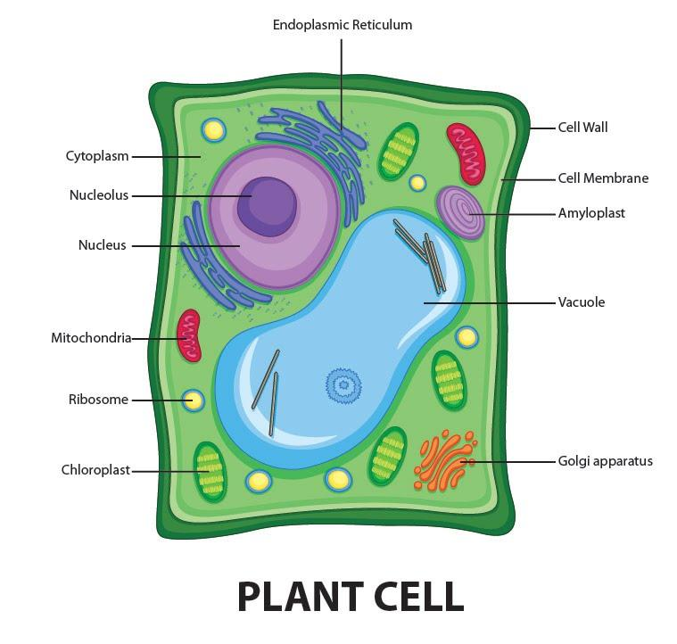
A plant cell contains a large, singular vacuole that is used for storage and maintaining the shape of the cell. In contrast, animal cells have many, smaller vacuoles. Plant cells have a cell wall, as well as a cell membrane. In plants, the cell wall surrounds the cell membrane. This gives the plant cell its unique rectangular shape.

Animal Cell Diagrams Labeled Printable 101 Diagrams
The plasma (cell) membrane separates the inner environment of a cell from the extracellular fluid. It is composed of a fluid phospholipid bilayer (two layers of phospholipids) as shown in figure 4.1.2 4.1. 2 below, and other molecules. Not many substances can cross the phospholipid bilayer, so it serves to separate the inside of the cell from.
Labeled Drawing Labeled Plant Cell And Animal Cell Diagram Goimages Fun
Cell diagram labeled Cell diagram unlabeled Learn faster with interactive cell quizzes Sources + Show all What are the parts of a cell? There exist two general classes of cells: Prokaryotic cells: Simple, self-sustaining cells (bacteria and archaea) Eukaryotic cells: Complex, non self-sustaining cells (found in animals, plants, algae and fungi)

What is a cell? Facts
There are six animal cell diagrams to choose from. The first is a colored and labeled cell diagram. The next is a black and white version of the first. These printables a free for subscribing members of Tim's Printables. Already a member? Please remember to log in. Not yet a member? Join today!
Animal cell hires stock photography and images Alamy
Key points: All cells have a cell membrane that separates the inside and the outside of the cell, and controls what goes in and comes out. The cell membrane surrounds a cell's cytoplasm, which is a jelly-like substance containing the cell's parts. Cells contain parts called organelles. Each organelle carries out a specific function in the cell.

identify and label each part of the eukaryotic cell
Cell organelles are specialized entities present inside a particular type of cell that performs a specific function. There are various cell organelles, out of which, some are common in most types of cells like cell membranes, nucleus, and cytoplasm. However, some organelles are specific to one particular type of cell-like plastids and cell.
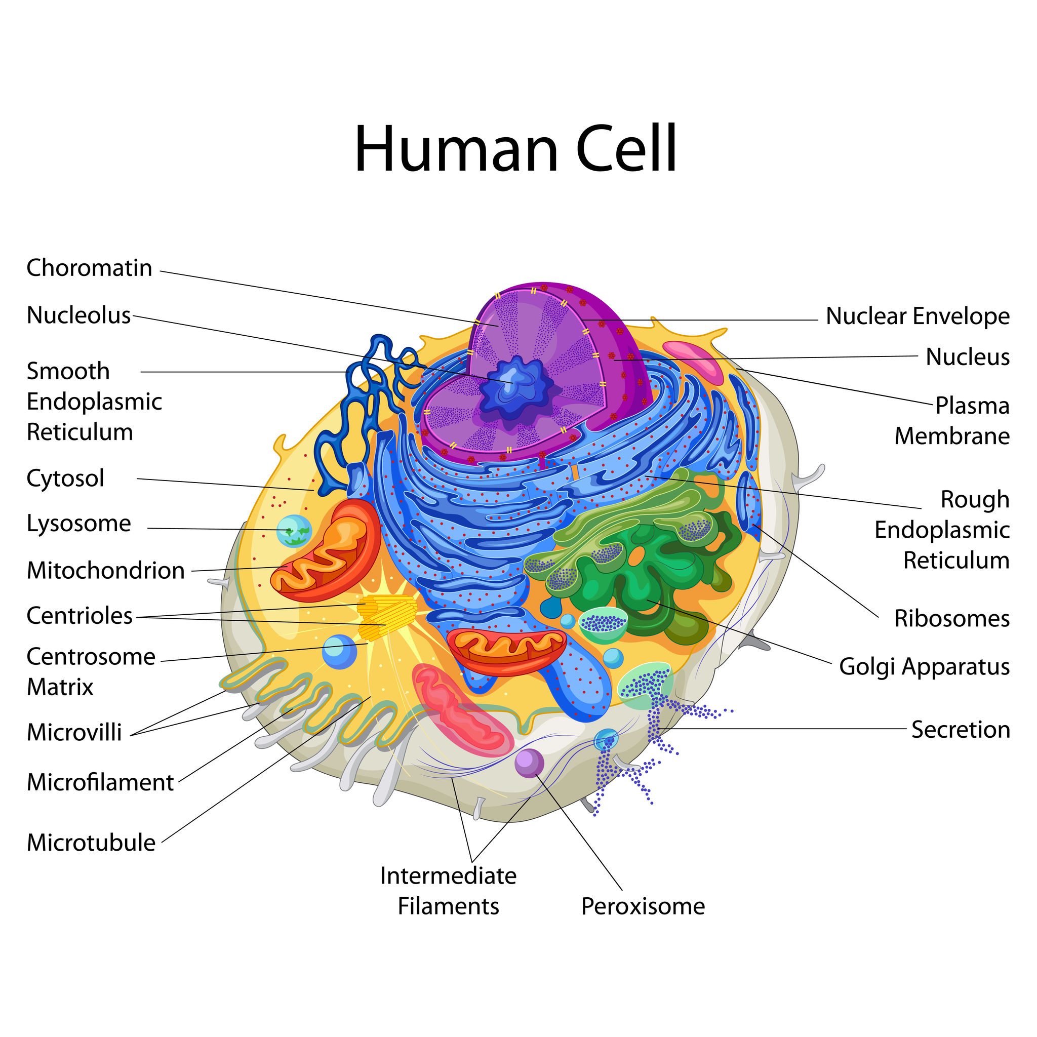
Education Chart of Biology for Human Cell Diagram Best Acupuncture llc
The cell structure illustrations for these diagrams were generated in BioRender. Both diagrams feature a drag-and-drop labelling activity created with H5P here on Learnful. These h5p resources are made available openly with the CC BY license. Plant Cell Structure: Animal Cell Structure:

Cells
Diagram Of Animal Cell Animal cells are eukaryotic cells that contain a membrane-bound nucleus. They are different from plant cells in that they do contain cell walls and chloroplast. The animal cell diagram is widely asked in Class 10 and 12 examinations and is beneficial to understand the structure and functions of an animal.

Pin on Animal cell
The nucleus is a large organelle that contains the cell's genetic information. Most cells have only one nucleus, but some have more than one, and others—like mature red blood cells—don't have one at all. Within the nucleus is a spherical body known as the nucleolus, which contains clusters of protein, DNA, and RNA.